Private Musculoskeletal Radiology Services
Diagnostics Services
Diagnostics
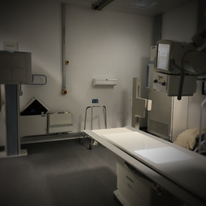
X-ray
X-rays are often the first-line investigation for demonstrating bone and joint abnormalities. The use of X-rays exposes the patient to a very small dose of radiation to produce an image in which all the anatomical structures are superimposed onto one another. Frequently, a radiographic examination requires two separate X-rays to be obtained at right angles. X-rays are produced in a digital format, which that can be viewed on a computer. While X-rays show exquisite bony detail soft-tissue features are poorly defined, and so other imaging techniques may be used to demonstrate soft-tissue abnormality.
Ultrasound
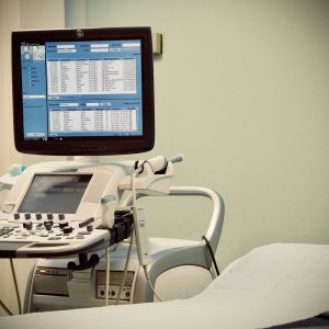
Ultrasound imaging is a common diagnostic medical procedure that uses painless high-frequency sound waves to produce dynamic pictures of organs, tissues and blood flow inside the body. Detailed images of your muscles, tendons and ligaments can be produced. The scan involves a hand-held probe that is placed directly onto the skin and moved over the patient. A water-based gel is used to couple the ultrasound between the probe and patient. Ultrasound can also be used to guide precise needle placement and treatment to areas of damaged tissue. The procedure takes 15-30 minutes.
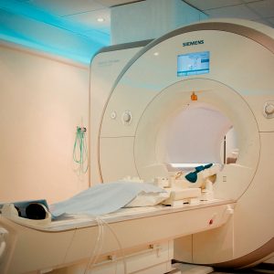
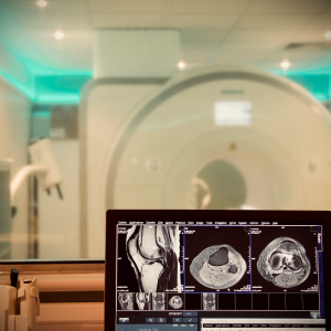
Magnetic Resonance Imaging (MRI)
MRI is a non-radioactive scan to show injury or disease in joint structures, such as muscles and ligaments, using strong magnetic and radio waves.
Occasionally, a contrast dye injection into the vein may be required to enhance the images. Please inform your doctor and the radiographer prior to your appointment if you have a pacemaker, metallic implant or have ever had a kidney condition.
The scan takes between 30 and 60 minutes.
Arthrograms
Arthrogram is an imaging process that involves the injection of contrast directly into the joint. The joint becomes enlarged by the procedure, which enhances the imaging of the smaller structures. This improves the evaluation of diseases or conditions of the joint. The procedure takes 15-30 minutes.
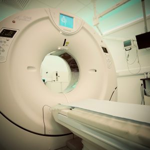
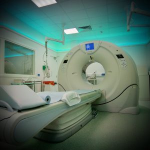
Computed Tomography (CT)
A CT scan is a painless scan that uses a special X-ray machine to take fine-detailed images of bones and joints. The scanner revolves around the body taking images as you lie on a moving table. Iodine-based contrast may be injected directing into the vein to enhance images. The technique uses a higher radiation dose than X-rays, but with modern equipment this is kept to a low level. The scan takes between 5 and 15 minutes.
As the procedure uses radiation, please inform a member of staff prior to your appointment if there is a chance you could be pregnant.
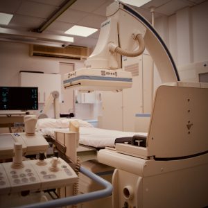
Fluoroscopy
Fluoroscopy is a means of producing real-time moving X-ray pictures by using a pulsed X-ray beam and displaying the images in real time on a TV monitor. Fluoroscopy can be utilised to ensure correct needle position for joint injections and interventional procedures.
CT
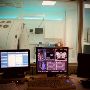
CT Control Room
Ultrasound
MRI
CT
MRI Control Room
Fluoroscopy
X-Ray
Interventional Services
Intervention
Image Guided Steroid Injections
Image Guided Steroid Injections
BMSR perform a wide range of cutting-edge, image guided injections. These procedures are carried out using specialist imaging equipment (X-ray, ultrasound or CT) which enable precise needle tip placement to the target tissue of interest e.g. joint, bursa, tendon or nerve sheath. Performing these injections under image guidance also allows for safe placement of the needle, avoiding unnecessary injury to adjacent structures e.g. blood vessels and nerves.
What is an image guided steroid injection?
It is an injection of local anaesthetic and steroid into the joints or soft tissues of your body. The local anaesthetic gives rapid pain relief. The steroid acts as a strong but localised anti-inflammatory to reduce pain and swelling, the effects of which last longer than the local anaesthetic.
Patients are referred for image guided injections for two reasons: Firstly, as a diagnostic test to confirm whether the pain or other symptoms that they are experiencing are arising from a particular joint, tendon or other structure. This can help guide any further investigations or treatment. Secondly as a therapeutic measure to help improve the pain or other symptoms.
What happens during the procedure?
The BMSR Consultant will explain the procedure to you and will answer any questions that you have.
You will be positioned in a chair or on a plinth or X-ray table.
Your skin will be cleaned with a sterile solution which will feel cold.
A local anaesthetic will be used to numb the area. This will feel sharp and sting for a few seconds. The injection needle will then be guided into the area of interest under x-ray, ultrasound or CT guidance. Sometimes, when using X-ray guidance, a contrast medium (dye) is used to confirm the needle tip position. Once the needle is correctly sited, a mixture of local anaesthetic and steroid is injected. The needle is the removed and a dressing or plaster applied.
Following the injection, it can be useful to document your response to the injection so that you can inform the specialist who referred you. (A pain diary will be given to you).
We suggest that you do not drive or operate machinery for the rest of the day. It is generally a good idea to take things easy for a few days following the injection.
As with all needle-stick interventions there are certain precautions, risks and complications which your BMSR Consultant will explain to you before carrying out the treatment (Read more on precautions, risks and complications).
Some commonly performed sites for guided injections include the following:
CT or X-ray guided:
- facet joint injections of the spine
- nerve root blocks and epidurals
X-ray guided:
- shoulder Hydrodilatation
- joint injection
Ultrasound guided:
- Shoulder – subacromial subdeltoid bursa
- Elbow – joint injection
- Wrist and hand – joints and tendon sheath
- Knee – joint, popliteal cyst, pes anserine bursa
- Ankle and foot – joints and tendon sheath, plantar fasciitis, Morton’s neuroma
Arthrograms
A joint arthrogram is an imaging procedure to investigate the spaces of a joint by injecting an X-ray dye into the joint before using an MRI scanner to take pictures.
The procedure is done under a local anaesthetic. However, you are advised not to drive after the procedure, so please make arrangements for someone to collect you from your appointment. Please allow an hour for the procedure.
The procedure uses X-rays; therefore, please inform a member of staff prior to your appointment if there is a chance you could be pregnant.
You will need to provide the department with a list of your current medications prior to your appointment. Please note, if you are taking blood thinners and do not inform the department before your appointment, your procedure may have to be cancelled.
Your BMSR Consultant will give you post procedure care advice at your appointment but please be aware that you will need to rest the joint for 12-24 hours.
Nerve Root Block
Nerve Root Block Injection
What is a nerve root block injection?
This is an injection around the nerve root as it leaves the spine. The injection is performed under image guidance (X-ray or CT)
Why do I need to have the injection?
The injection is usually done to relieve pain and inflammation around the nerve. It is used to help arm or leg pain (sciatica) and paraesthesia (pins and needles) and is sometimes used for back pain. The injection may also be useful to help diagnose the exact source of your pain.
What is injected?
- An anti-inflammatory drug (steroid).
- A local anaesthetic.
- A small quantity of dye is also injected so that the BMSR Consultant Radiologist can be sure that the injection is around the nerve
The local anaesthetic will be responsible for any immediate relief of symptoms. When this wears off the pain may return before the benefits of the anti-inflammatory is felt. The anti-inflammatory drug may take up to 6 weeks to work. Pain may return after some time. If this occurs a decision by your referring doctor will then be made about repeating the nerve root injection or considering other possible treatments.
Precautions
Please inform BMSR if you;
- are pregnant
- have diabetes
- feel unwell
- have an infection, cold or persistent cough
- have any allergies
- are taking any of the following medications – antibiotics, asprin, warfarin or clopidrogrel or any other tablets used to thin the blood (some of these may need to be stopped some days before). Failure to do so may result in the procedure being cancelled.
What are the risks of this procedure?
Please note the risks from having a nerve root block are small.
- Allergic reaction to the injection
- Infection
- Damage to the small veins on insertion of the needle
- Bleeding causing local bruising or bleeding around the nerve
- Nerve damage
- If you have diabetes then the injection may raise your glucose levels. These should be monitored for up to 1 month after the injection. If there are any changes in your diabetic symptoms, please consult your GP
- Facial flushing for a few days
- Temporary discomfort for a few days after your injection
- For females – temporary alteration of the menstrual cycle
What the procedure involves
You will be lying on your front for the procedure, which usually takes 10-20 minutes. Local anaesthetic is injected into the skin and a fine needle is passed towards the nerve root under X-ray guidance. Once the needle is confirmed to be close to the nerve, the injection takes place.
What happens after your procedure
You will be monitored by the nursing staff on the unit until you are ready to go home (about 30 minutes).
Please note: You must not drive yourself home or use public transport due to a potential weak leg if you are having a lumbar nerve root block.
For your own well-being, we advise that you are collected by a relative or friend.
Back at home
It is important that you take things easy for the rest of the day. Do not do any excessive exercise or heavy work for the first few days. If a dressing is in place, remove the dressing the morning following your procedure. Continue to take your painkiller medication until you notice an improvement in your symptoms.
Follow up appointment
The need for a follow-up appointment will be discussed before you are discharged.
Facet Joint Injections for Back Pain
Facet joint injections are used to treat pain from degenerative changes within the spine, which can be one of the many causes of backache. Steroid and a small amount of local anaesthetic can be injected into the small joints under X-ray guidance to give pain relief. Speak to your medical professional to discuss whether these injections can help your pain.
The procedure can take 30 minutes.
You should stay within the department for 15-30 minutes after the procedure and are advised not to drive yourself home.
Hydrodilatation for Frozen Shoulder
What is frozen shoulder?
Frozen shoulder (also known as adhesive capsulitis) is an extremely painful condition in which the shoulder becomes very stiff (hence the term ‘frozen’). In most cases frozen shoulder starts out of the blue for no apparent reason. However, it may be associated with other conditions such as diabetes, thyroid disease or Dupuytren’s disease. It may also be triggered by a mild injury or surgery to the shoulder. The condition usually goes through three distinct phases, starting with pain (the freezing stage), then stiffness (the frozen stage) and finally a stage of resolution as the pain eases and most of the movement returns (the thawing phase). This process may take a long time, sometimes as long as 2 years or more.
The lining of the shoulder joint, known as the ‘capsule’, is normally a very loose elastic structure – this allows for the large range of motion and flexibility of the joint. With a frozen shoulder the capsule of the shoulder joint becomes inflamed, swollen and contracted. The end result is a painful and stiff joint.
Hydrodilatation of the shoulder
A fine needle is inserted into the shoulder joint under X-ray guidance (sometimes this can also be performed under ultrasound guidance). Then, 15-40 mls of a mixture containing normal saline (salty water), local anaesthetic and a small amount of steroid is then injected into the shoulder joint. A small amount of dye is first injected to ensure correct positioning of the needle tip.
The fluid distends and ‘stretches out’ the contracted capsule of the shoulder joint thereby opening or freeing up the joint and allowing an improved range of movement. The steroid helps to ease with the pain and inflammation.
The procedure can sometimes be uncomfortable (patients will frequently describe a pressure build up within the shoulder), although this is not always the case. However, most patients will notice an immediate reduction in their pain and an increased range of motion following the procedure.
It is generally a good idea to perform remedial exercises of the shoulder following the procedure in order to further improve your symptoms.
Shoulder hydrodilatation is a safe and minimally invasive procedure. However, as with all needle-stick interventions there are certain precautions, risks and complications which your BMSR Consultant will explain to you before carrying out the treatment.
Barbotage Therapy
Image-guided barbotage is a procedure used to break up calcification within tendons. The commonest location of calcification is within the shoulder tendons (calcific tendinosis). Under local anaesthetic and ultrasound guidance, a needle is used to break up and suck out the calcium. Steroid is then injected to reduce inflammation. The procedure can take up to 30 minutes.
You should stay within the department for 15-30 minutes after the procedure and are advised not to drive yourself home.
As the local anaesthetic wears off, it is advisable to continue taking your painkiller medication as the steroid can take several days to work
Brisement Therapy
Image-guided brisement therapy is used to break up scar tissue and is commonly performed for painful inflammation of the Achilles tendon. It is a high-volume injection of local anaesthetic and saline, which is injected under ultrasound guidance. The procedure can take 15-30 minutes.
You should stay within the department for 15-30 minutes after the procedure and are advised not to drive yourself home.